
Research
Applied Spectroscopy

Our research in spectroscopy involves using the interaction of light with matter to identify and quantify components within a sample. We address a variety of critical real-world challenges. For example, we’re using these methods for authenticating whisky and other alcohols in sealed bottles, tackling antimicrobial resistance, assess the metabolic activity of developing embryos, and identifying hazardous pigments in old books.
Recent Publications in Applied Spectroscopy:
- Detecting emerald green in 19th century books using light, STEM for Britain, Houses of Parliament, Westminster, (2025).
- Monitoring live mycobacteria in real-time using a microfluidic acoustic-Raman platform, Antibiotic Resistance Protocols 2833, 109 (2024).
- Learning algorithms for identification of whisky using portable Raman spectroscopy, Current Research in Food Science 8, 100729 (2024).
- UVA hyperspectral light-sheet microscopy for volumetric metabolic imaging: application to pre-implantation embryo development, ACS Photonics 10, 4177 (2023).
- To focus-match or not to focus-match inverse spatially offset Raman spectroscopy: a question of light penetration, Opt. Express 30, 8876 (2022).
- Through-bottle whisky sensing and classification using Raman spectroscopy in an axicon-based backscattering configuration, Anal. Methods 12, 4572 (2020).
Imaging and Microscopy
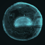
By precisely controlling the wavefront of light, we can image biomedical objects such as tissue and developing embryos, at high resolution while significantly reducing the required light exposure. This translates to less damage to delicate biological samples and faster image acquisition. Our techniques allow us to see through highly scattering materials like biological tissue, and we are also developing cutting-edge hyperspectral methods that enable us to understand both the chemical composition and morphological features within complex images.
Recent Publications in Imaging and Microscopy:
- Estimating full-field displacement in biological images using deep learning, npj Artificial Intelligence 1, 6 (2025).
- Optically generated droplet beams improve optoacoustic imaging of choroid thickness as an Alzheimer’s disease biomarker, npj Nanophotonics 1, 42 (2024).
- Sidelobe suppressed Bessel beams for one-photon light-sheet microscopy, Biomed. Opt. Express 15, 6183 (2024).
- UVA hyperspectral light-sheet microscopy for volumetric metabolic imaging: application to pre-implantation embryo development, ACS Photonics 10, 4177 (2023).
- Spatially offset optical coherence tomography: leveraging multiple scattering for high-contrast imaging at depth in turbid media, Sci Adv. 9, eeadh5435 (2023).
- Investigation of refractive index dynamics during in vitro embryo development using off-axis digital holographic microscopy, Biomed. Opt. Express 14, 3327 (2023).
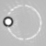
Speckle Metrology

Speckle metrology is a powerful technique that utilizes the random interference patterns (speckles) formed when coherent light illuminates a rough surface. Our group harnesses these patterns to perform highly resolved measurements of properties of light or the environment, such as attometer-resolved wavelength measurements, picometer-resolved displacement measurements and measurements of refractive index changes with <1 ppm sensitivity.
Recent Publications in Speckle Metrology:
- Determining intrinsic sensitivity and the role of multiple scattering in speckle metrology, Nature Rev. Phys. 6, 500 (2024).
- Measuring picometre-level displacements using speckle patterns produced by an integrating sphere, Sci. Rep. 13, 14607 (2023).
- Measurement of variations in gas refractive index with 10−9 resolution using laser speckle, ACS Photonics 9, 830 (2022).
- Wavelength sensitivity of the speckle patterns produced by an integrating sphere, J. Phys. Photonics 3, 035005 (2021).
- Speckle-based determination of the polarisation state of single and multiple laser beams, OSA Continuum 3, 1302 (2020).
- Overcoming the speckle correlation limit to achieve a fiber wavemeter with attometer resolution, Opt. Lett. 44, 1367 (2019).
Optical Trapping and Manipulation
A strongly focused beam of light can be used to trap and move objects ranging in size from tens of nanometers to tens of micrometers. These optical tweezers have become a widely-used tool in biology, physical chemistry, and fundamental physics and can be harnessed in both liquid and vacuum environments. Our group has made significant recent contributions in the areas of Optically Induced Rotation, Non-Reciprocal Forces, and Optical Binding.
Recent Publications in Optical Trapping:
- Microrheology of the cumulus-oocyte matrix using optical tweezers, J. Phys. Photonics 7, 025008 (2025).
- Fano resonance-assisted all-dielectric array for enhanced near-field optical trapping of nanoparticles, ACS Photonics 10, 4322 (2023).
- Roadmap for optical tweezers, J. Phys. Photonics 25, 022501 (2023).
- Cooling the optical-spin driven limit cycle oscillations of a levitated gyroscope, Commun. Phys. 6, 238 (2023).
- Laser writing of parabolic micromirrors with a high numerical aperture for optical trapping and rotation, Appl. Phys. Lett. 123, 081106 (2023).
- All-optical sub-kelvin sympathetic cooling of a levitated microsphere in vacuum, Optica 9, 1000 (2022).