
Optical Imaging
New ways of imaging with shaped light can lead us to take images of large biomedical objects (eg tissue, small organs) at high resolution with minimal exposure to light, meaning less damage and faster image acquisition, and allow us to see through scattering material such as tissue.
Light Sheet Microscopy
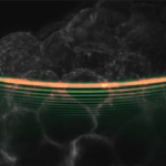
In light sheet microscopy, we illuminate samples with a broad sheet of light rather than point by point scanning. This leads to faster image acquisition and lower sample exposure, thus leading to less “light damage”. Such imaging can lead to new insights for studies in neuroscience, diseases of the mind (dementia) and developmental biology. In turn this may shed light on numerous biological processes including the development of disease. In particular, we harness the non-diffracting properties of Airy beams in our light sheet microscopes to increase the field of view deeper into the sample.
- WIDEFIELD LIGHT SHEET MICROSCOPY USING AN AIRY BEAM COMBINED WITH DEEP-LEARNING SUPER-RESOLUTION
S. Corsetti, P. Wijesinghe, P. B. Poulton, S. Sakata, K. Vyas, C. S. Herrington, J. Nylk, F. Gasparoli and K. Dholakia, OSA Continuum 3, 1068-1083 (2020) - LIGHT SHEET FLUORESCENCE MICROSCOPY FOR NEUROSCIENCE
S. Corsetti, F. J. Gunn-Moore and K. Dholakia, Journal of Neuroscience Methods 319, 16 (2019) - LIGHT SHEET MICROSCOPY WITH ACOUSTIC SAMPLE CONFINEMENT
Z. Yang, K. L. Cole, Y. Qiu, I. M. L. Somorjai, P. Wijesinghe, J. Nylk, S. Cochran, G. C. Spalding, D. A. Lyons, and K. Dholakia, Nat. Commun. 10, 669 (2019) - THREE-PHOTON LIGHT-SHEET FLUORESCENCE MICROSCOPY
A. Escobet-Montalbán, F. M. Gasparoli, J. Nylk, P. Liu, Z. Yang and K. Dholakia, Opt. Lett. 43, 5484 (2018) - LIGHT-SHEET MICROSCOPY WITH ATTENUATION-COMPENSATED PROPAGATION-INVARIANT BEAMS
J. Nylk, K. McCluskey, M. A. Preciado, M. Mazilu, Z. Yang, F. J. Gunn-Moore, S. Aggarwal, J. A. Tello, D. E. K. Ferrier and K. Dholakia, Science Advances 4, eaar4817 (2018) - LIGHT-SHEET MICROSCOPY USING AN AIRY BEAM
T. Vettenburg, H. I. C. Dalgarno, J. Nylk, C. Coll-Lladó, D. E. K. Ferrier, T. Čižmár, F. J. Gunn-Moore and K. Dholakia , Nature Methods 11, 541-544 (2014)
TRAFIX Microscopy
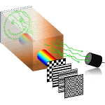
The light-scattering properties of tissue make it incredibly hard to record images from deep within biological samples. We approach this challenge by implementing a scheme that requires no a priori information about the medium nor its properties. Exploiting temporal focusing, patterned illumination and single-pixel detection in our innovative scheme, we obtain wide-field two-photon images from deep within samples. The illumination intensity can be reduced further by optimising the basis in which the sequential illumination patterns are encoded.
- OPTIMAL COMPRESSIVE MULTIPHOTON IMAGING AT DEPTH USING SINGLE-PIXEL DETECTION
P. Wijesinghe, A. Escobet-Montalbán, M. Chen, P. R. T. Munro and K. Dholakia, Opt. Lett. 44, 4981 (2019) - WIDE-FIELD MULTIPHOTON IMAGING THROUGH SCATTERING MEDIA WITHOUT CORRECTION
A. Escobet-Montalbán, R. Spesyvtsev, M. Chen, W. Afshar Saber, M. Andrews, C. S. Herrington, M. Mazilu and K. Dholakia, Science Advances 4, eaau1338 (2018) - WIDE-FIELD 3D OPTICAL IMAGING USING TEMPORAL FOCUSING FOR HOLOGRAPHICALLY TRAPPED MICROPARTICLES
R. Spesyvtsev, H. Rendall and K. Dholakia, Opt. Lett. 40, 4847 (2015)