
Sound and vision – the future of drug discovery
A marriage of sound and light could hold the key to diagnosis of the early stages of various diseases, including cancer, new research led by the University of St Andrews reveals.
The international research team, in collaboration with the University of Edinburgh, University of Glasgow, Illinois Wesleyan University, USA, and the University of Western Australia, Perth, developed an innovative new way to hold samples using sound whilst they are gently imaged using light.
Published in the journal Nature Communications, the research paves the way to move to a host of new studies for advanced drug discovery and combating conditions such as coronary heart disease.
The ability to hold and image objects has had a profound impact across biomedical sciences, especially those focused upon disease identification, neuroscience and developmental biology. The innovative approach developed by the team uses sound waves to grip small samples. The sound wave or acoustic beam impinging on an object can result in a “radiation force” as the object scatters the sound waves. If the beam is suitably shaped, this force can be used to immobilise an object. The normal way to immobilise the object would be to use a gel but is not compatible with many organisms. More pertinently, when one wants to introduce drugs to the sample this can be slow and unpredictable as they have to permeate through pores in the gel. The use of sound solely to immobilise samples removes this issue as the object is held in its native liquid medium.
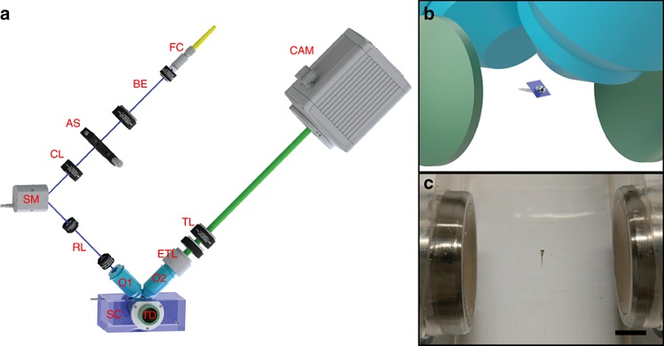
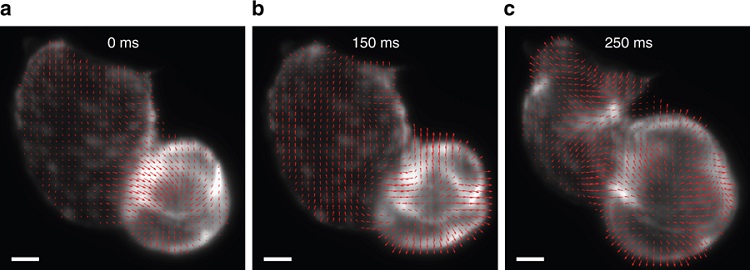
The team held a range of samples including zebrafish embryos and larvae and a variety of marine animals. Once held, the sample was rapidly sampled by light sheet imaging, a powerful way to rapidly acquire three dimensional volumes of organisms.
As an example, they monitored the reaction of the heartbeat of zebrafish larvae to drugs, which are known to affect cardiovascular function. They explored the cardiac cycle of the zebrafish and described a new way to see changes in contractile volume within the heart that in future could lead to new insights for cardiovascular research
Professor Kishan Dholakia, from the University of St Andrews School of Physics and Astronomy, said: “This is a new way to perform drug-based studies for cardiovascular disease and developmental biology. We anticipate this approach can also be used for high throughput drug discovery, an important topic for future healthcare.”
The paper Light sheet microscopy with acoustic sample confinement is published in Nature Communications and is available online.
The research was supported by the UK Engineering and Physical Sciences Research Council, the European Union’s Horizon 2020 Programme through the project Advanced BiomEdical OPTICAL Imaging and Data Analysis (BE-OPTICAL), The Cunningham Trust and The RS MacDonald Charitable Trust, the Wellcome Trust ISSF, and the EU Horizon 2020 Programme. The University of St Andrews has protected the intellectual property rights by filing a patent application.